Regular X-ray machines and CT scanners can produce 2-D or 3-D images but in monochrome. In the same way that black-and-white film is unsighted to other wavelengths of light. These techniques cannot distinguish between different wavelengths of X-ray whereas the colour X-ray machine is able to distinguish between different wavelengths. Pathological soft tissue specimens are expected to show a small contrast in absorption whereas diffraction patterns will be significantly different between healthy and diseased tissue.
©Shutterstock
New Zealand scientist performs first ever 3-D, colour X-ray on a human using a technique called TEDDI. This promises to improve the field of medical diagnostics.
How the colour X-ray machine works
After a sample is hit with an X-ray beam, the device collects the scattered X-rays onto the different pixels of the detector. Each looks at one area of the sample, you move the sample through the scanner to get a full 3D image. This new technique is called Tomographic Energy Dispersive Diffraction Imaging, or TEDDI.”This colour X-ray imaging technique could produce clearer and more accurate pictures and help doctors give their patients more accurate diagnoses,” said a CERN statement. In this new technology known as the spectral CT, the sensor can measure the attenuation of specific wavelengths of the X-rays as they pass through different materials. After running the spectroscopic data through specific algorithms, a 3D colour image is generated. This image clearly shows muscle, bone, water, fat, disease markers – and even a watch. The end results are unnerving like someone’s sculpted a detailed clay model of your insides.
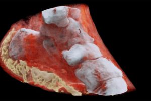
Below are the images of the World’s first ever colour X-ray performed on the human body
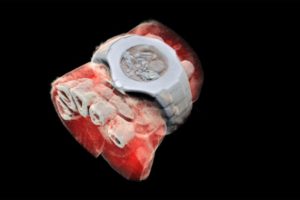
A 3D image of a wrist with a watch showing part of the finger bones in white and soft tissue in red © HO / MARS Bioimaging Ltd / AFP
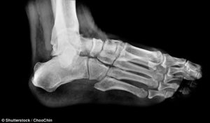
A 3D image of the left view of an ankle, with its bones in white and soft tissue in red.© HO / MARS Bioimaging Ltd / AFP
These images give an improved visualisation regarding the diagnosis without cutting you open. According to the CERN, the images very clearly show the difference between bone, muscle and cartilage. In addition, they also show the position and size of cancerous tumours. Presently the technology is being commercialised by New Zealand company MARS Bioimaging. It is also linked to the universities of Otago and Canterbury which helped develop it.


1 Comment
elly camron · August 4, 2021 at 11:36 am
This content is very helpful for me. Thanks for sharing